MSK Anatomy Masterclass
A comprehensive bilingual deep-dive into musculoskeletal anatomy — joints, muscles, ligaments, and clinical relevance. Includes anatomical diagrams, imaging interpretation, and real case-based learning across 12 modules.
4.9
Rating
320+
Students
6h
Content
65
Lessons
Afonso Vera
MSc Physiotherapy (MMU) · BSc Sport Rehab (Salford) · 5yr MSK · 8yr Rugby
£49
One-time payment · Lifetime access
30-day money-back guarantee
What You Will Learn
Identify and describe key bony landmarks, joints, muscles, ligaments & neurovascular structures of all major MSK regions
Explain the biomechanics and functional anatomy relevant to clinical assessment
Apply anatomical knowledge to interpret common MSK presentations and pathologies
Perform confident surface anatomy palpation using correct anatomical terminology
Integrate anatomy with clinical reasoning for differential diagnosis
Interpret basic MSK imaging (X-ray, MRI, ultrasound) and correlate with clinical findings
Course Curriculum
12 modules · 65 lessons · 6 hours total
Objective: Establish the anatomical language and frameworks used throughout the course.
Anatomical planes, directions & terminology (EN + PT glossary)
Bone types, joint classifications & connective tissue overview
How to use anatomy in clinical reasoning — the physio's approach
Surface anatomy principles: palpation landmarks & technique
Key concepts: Sagittal/coronal/transverse planes, synovial joints, cartilage types, enthesis, proprioception
Objective: Understand the anatomy of the cervical spine and its clinical relevance to neck pain and radiculopathy.
Bony anatomy: C1–C7 vertebrae, facet joints, uncovertebral joints
Intervertebral discs and the cervical lordosis
Muscles: deep neck flexors, extensors, and lateral flexors
Neurovascular anatomy: brachial plexus roots, vertebral artery
Clinical case: Cervical radiculopathy — anatomy behind the symptoms
Key concepts: Atlanto-axial joint, deep cervical flexors, C5–C6 dermatomes, Spurling's test anatomy
Objective: Master lumbar and thoracic anatomy with direct application to low back pain assessment.
Thoracic vertebrae, rib articulations & costovertebral joints
Lumbar spine: L1–L5 anatomy, facet joint orientation & movement
The intervertebral disc: nucleus pulposus, annulus fibrosus & herniation mechanics
Paraspinal muscles: erector spinae, multifidus, quadratus lumborum
The lumbar plexus and sciatic nerve pathway
Clinical case: Lumbar disc herniation with L4/L5 radiculopathy
Key concepts: Multifidus, thoracolumbar fascia, SIJ anatomy, sciatic nerve, straight leg raise anatomy
Objective: Develop a thorough understanding of the shoulder as a functional unit across four joints.
The four joints: glenohumeral, acromioclavicular, sternoclavicular, scapulothoracic
Rotator cuff: supraspinatus, infraspinatus, teres minor, subscapularis
The glenoid labrum, joint capsule & glenohumeral ligaments
Scapular stabilisers: serratus anterior, trapezius, rhomboids
Neurovascular anatomy: brachial plexus, axillary nerve, suprascapular nerve
Clinical case: Rotator cuff tear — anatomy of impingement & full-thickness tears
Key concepts: Subacromial space, scapulohumeral rhythm, GIRD, Hawkins-Kennedy anatomy, axillary nerve at risk
Objective: Understand elbow joint anatomy and its relevance to lateral/medial epicondylalgia and UCL injuries.
Bony anatomy: humeroulnar, humeroradial & proximal radioulnar joints
Medial & lateral ligament complexes: UCL, RCL, annular ligament
Muscles: flexors, extensors & the common extensor/flexor origins
Neurovascular anatomy: radial nerve, ulnar nerve at cubital tunnel, median nerve
Clinical case: Lateral epicondylalgia — anatomy of the ECRB & tendinopathy
Key concepts: Carrying angle, cubital tunnel, radial tunnel, common extensor origin, UCL in throwing athletes
Objective: Navigate the complex anatomy of the wrist and hand relevant to common MSK presentations.
Carpal bones, radiocarpal joint & intercarpal joints
Intrinsic & extrinsic hand muscles: thenar, hypothenar, lumbricals, interossei
Flexor & extensor tendons: pulleys, sheaths & zones
Neurovascular anatomy: carpal tunnel, Guyon's canal, digital nerves
Clinical case: De Quervain's tenosynovitis — anatomy of the first extensor compartment
Key concepts: Scaphoid blood supply, TFCC, carpal tunnel contents, flexor pulley system
Objective: Understand hip joint anatomy and pelvic biomechanics in the context of hip pain and FAI.
Bony anatomy: acetabulum, femoral head, neck-shaft angle, anteversion
Hip joint capsule, labrum & ligamentous anatomy
Hip muscles: gluteal group, hip flexors, adductors, external rotators
Pelvic floor anatomy & its relationship to lumbopelvic stability
Neurovascular anatomy: femoral nerve, sciatic nerve, lateral femoral cutaneous nerve
Clinical case: Femoroacetabular impingement (FAI) — cam vs pincer anatomy
Key concepts: Q-angle, greater trochanteric anatomy, piriformis & sciatic nerve, Thomas test anatomy, Trendelenburg anatomy
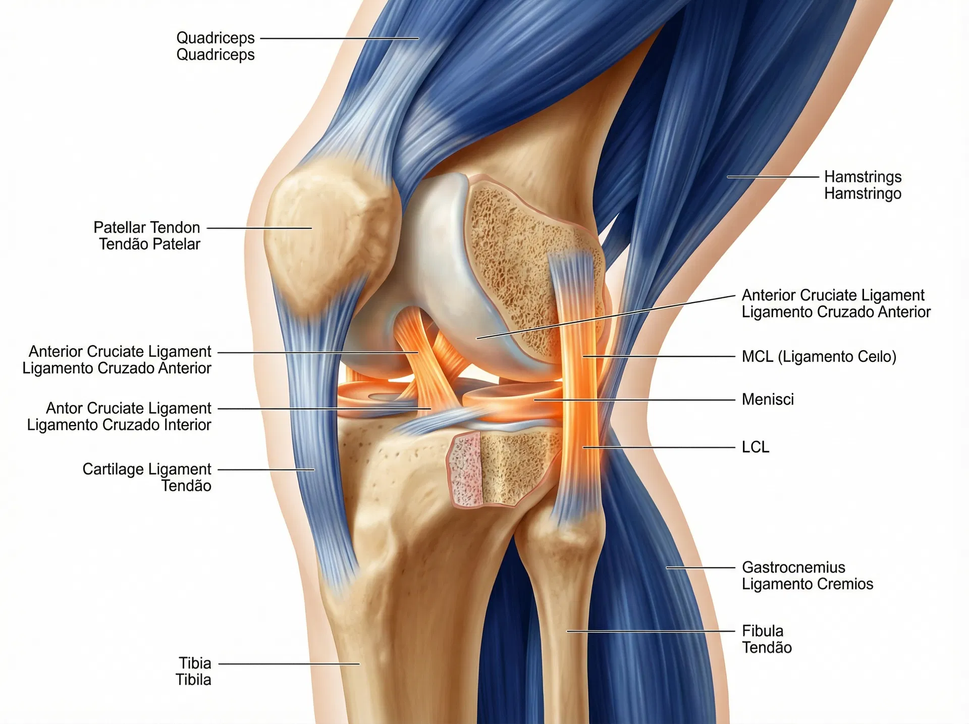
Objective: Achieve a thorough understanding of knee anatomy for the most common MSK joint in clinical practice.
Bony anatomy: tibiofemoral & patellofemoral joints, tibial plateau, femoral condyles
Cruciate ligaments: ACL & PCL — anatomy, fibre bundles & biomechanics
Medial & lateral structures: MCL, LCL, posterolateral corner
Menisci: medial vs lateral, blood supply zones & tear patterns
Extensor mechanism: quadriceps, patella, patellar tendon & MPFL
Neurovascular anatomy: popliteal fossa contents, common peroneal nerve
Clinical case: ACL rupture — anatomy of the injury & reconstruction graft options
Key concepts: Lachman's anatomy, pivot shift, meniscal blood supply zones, patellofemoral contact pressure
Objective: Understand ankle and foot anatomy relevant to sprains, Achilles pathology, and plantar fasciitis.
Bony anatomy: talocrural, subtalar, midtarsal & tarsometatarsal joints
Lateral ankle ligaments: ATFL, CFL, PTFL — anatomy & sprain grades
Medial deltoid ligament complex & syndesmosis anatomy
Muscles of the ankle & foot: extrinsic & intrinsic groups
Plantar fascia anatomy & the windlass mechanism
Clinical case: Achilles tendinopathy — anatomy of the tendon zones & vascularity
Key concepts: Sinus tarsi, tarsal tunnel, peroneal tendons, Achilles watershed zone, Köhler's fat pad
Objective: Apply peripheral neuroanatomy to clinical assessment and differential diagnosis.
Peripheral nerve structure: epineurium, perineurium, endoneurium
Dermatomes & myotomes: upper and lower limb clinical reference maps
Common nerve entrapment syndromes: carpal tunnel, cubital tunnel, tarsal tunnel, piriformis
Neural tension tests: anatomy behind ULNT1–4, SLR & slump test
Clinical case: Double crush syndrome — anatomy of multi-level nerve compression
Key concepts: Axonal transport, Sunderland classification, neural mechanosensitivity, adverse neural tension
Objective: Develop the ability to interpret basic MSK imaging and correlate findings with clinical presentation.
X-ray anatomy: reading joint spaces, bony alignment & degenerative changes
MRI anatomy: identifying muscles, tendons, ligaments & cartilage on T1/T2
Ultrasound anatomy: real-time tendon, muscle & bursa identification
Common imaging findings: disc bulge vs herniation, rotator cuff tears, meniscal tears
Clinical case: Correlating MRI findings with clinical examination
Key concepts: Modic changes, STIR sequences, hyperechoic vs hypoechoic tendons, incidental findings
Objective: Synthesise anatomical knowledge across all regions through three complex clinical cases.
Case A: The rugby player with multi-region pain — shoulder, lumbar spine & knee
Case B: The desk worker with cervical radiculopathy & carpal tunnel syndrome
Case C: The runner with hip, knee & ankle pain — a biomechanical anatomy chain
Building your anatomy-first clinical reasoning framework
Course summary, key takeaways & recommended further reading
Key concepts: Regional interdependence, kinetic chain anatomy, anatomy as the foundation of clinical reasoning
Who Is This Course For?
Physiotherapy students (BSc and MSc level)
Sport rehabilitation students and graduates
Newly qualified physiotherapists building clinical confidence
Coaches, athletic trainers & sports medicine practitioners
Portuguese-speaking clinicians seeking bilingual CPD
Anyone wanting to strengthen their anatomical foundation
£49
One-time payment · Lifetime access
30-day money-back guarantee
This course includes:
12 modules across 6 hours of content
Bilingual delivery: English and Portuguese throughout
Downloadable anatomy reference sheets for each region
2 clinical case studies per module
End-of-module knowledge check questions
Certificate of completion
Lifetime access with free updates
High-quality anatomical diagrams & 3D illustrations
Your Instructor
Afonso Vera
MSc Physiotherapist
MSc Pre-Reg Physiotherapy at Manchester Metropolitan University, BSc Sport Rehabilitation at the University of Salford, Sports Massage Level 3. 5 years MSK clinical experience and 8 years working in semi-professional rugby union in the UK. Bilingual EN/PT clinician and educator.
Ready to Master MSK Anatomy?
Join 320+ students who have already transformed their anatomical knowledge and clinical confidence.
One-time payment · Lifetime access · 30-day money-back guarantee